Drawing Of Esophagus
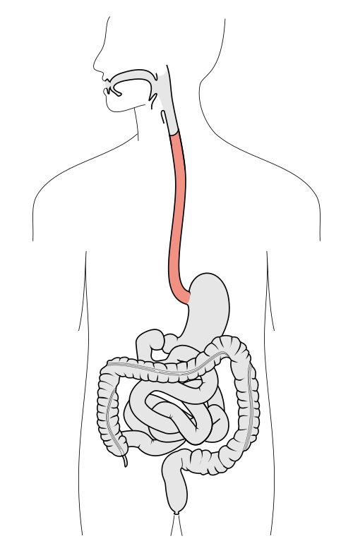
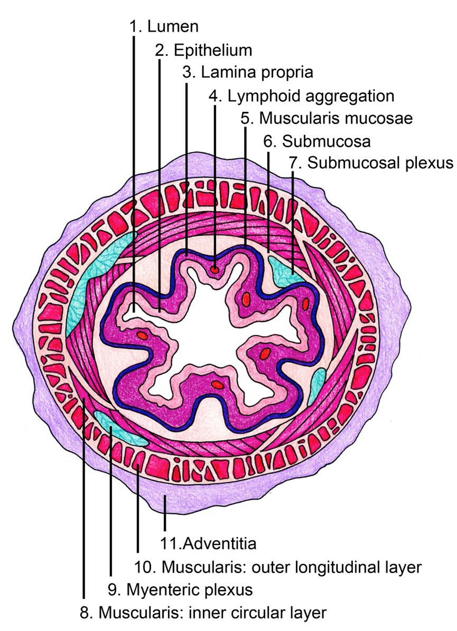
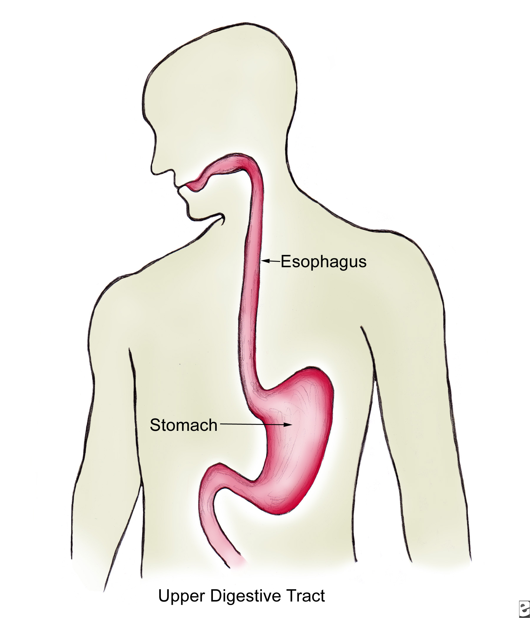
Drawing Of Esophagus - It passes through the diaphragm. This integration significantly increases the diagnostic accuracy for ev. Esophageal cancer ranks number six of the cancers that cause death. A pullout shows the mucosa layer, thin muscle layer, submucosa layer, thick muscle layer, and connective tissue layer of the esophagus wall. Web how to draw esophagus and mouth anotamy drawing The food moves from the mouth into the esophagus, which carries it down into the stomach. Web drawing inspiration from the thorough assessment practices of radiologists, moon establishes a cohesive multiorgan analysis model that unifies the imaging features of the related organs of ev, namely esophagus, liver, and spleen. The inner lining of the esophagus is a layer of soft tissue, called the mucosa (or innermost mucosa), is itself composed of three layers. Isolated vector illustration human gut. Drawing shows the pharynx (throat), esophagus, and stomach. Drawing shows the pharynx (throat), esophagus, and stomach. Web choose from esophagus drawing stock illustrations from istock. It passes through the diaphragm. 2 renmin hospital of wuhan university, wuhan, hubei province, china ; Web los angeles dodgers righty dustin may will miss the rest of this season after undergoing surgery to repair a torn esophagus, the team announced. Web the esophagus is the tube that connects the mouth and throat (pharynx) to the stomach. Web the esophagus is one of the upper parts of the digestive system.there are taste buds on its upper part. It consists of muscles that run both longitudinally and circularly, entering into the abdominal cavity via the right crus of the diaphragm at the level of the tenth thoracic vertebrae. When the patient is upright, the esophagus is usually between 25 to 30 centimeters. Web your esophagus is a hollow, muscular tube that carries food and liquid from your throat to your stomach. The esophagus is situated in front of the spine and behind the heart and trachea (windpipe). At the end of the mouth, draw a small tube that extends straight down into the center of your model’s torso. The esophagus is composed of four tunics (layers): Web 1 school of mathematics and economics, hubei university of education, wuhan, hubei 430205, china,. Mucosa > the mucosa of the esophagus is lined with stratified squamous moist epithelium to protect the organ from the partially. The lymph nodes are also shown. Isolated vector illustration human gut. The esophagus is a muscular tube about ten inches (25 cm.) long, extending from the hypopharynx to the stomach.the esophagus lies posterior to the trachea and the heart. Web the esophagus is a hollow muscular tube that transports saliva, liquids, and foods from the mouth to the stomach. It is located just posterior to the trachea in the neck and thoracic regions of the body and passes through the esophageal hiatus of the diaphragm on its way to the stomach. It consists of muscles that run both longitudinally. It is located just posterior to the trachea in the neck and thoracic regions of the body and passes through the esophageal hiatus of the diaphragm on its way to the stomach. The esophagus is situated in front of the spine and behind the heart and trachea (windpipe). Web the esophagus is one of the upper parts of the digestive. The inner lining of the esophagus is a layer of soft tissue, called the mucosa (or innermost mucosa), is itself composed of three layers. It passes through the diaphragm. It consists of muscles that run both longitudinally and circularly, entering into the abdominal cavity via the right crus of the diaphragm at the level of the tenth thoracic vertebrae. Web. 02:45layers of the esophageal wall: Problems with the esophagus include. Web content:introduction 0:00parts of the esophagus: Its exterior, the epithelium, is composed of protective cells, with layers of connective tissue (lamina propria) and thin bands of smooth muscle (muscularis mucosa). When these cells mutate and cause masses we see a cancer that is called squamous cell carcinoma. This is the most common type of esophageal cancer in the. The esophagus is composed of four tunics (layers): Web anatomy of the esophagus. It begins at the back of the mouth, passing downward through the rear part of the mediastinum, through the diaphragm, and into the stomach.in humans, the esophagus generally starts around the level of the sixth cervical. Here in this section i am going to share esophagus slide image. Do you want to get esophagus histology slide drawing tutorial? The camera is able to visualize the esophagus and stomach, and biopsies are obtained through the endoscope. The esophagus is made of smooth muscle that. Web los angeles dodgers righty dustin may will miss the rest of this. Here in this section i am going to share esophagus slide image. Web drawing inspiration from the thorough assessment practices of radiologists, moon establishes a cohesive multiorgan analysis model that unifies the imaging features of the related organs of ev, namely esophagus, liver, and spleen. The injury did not occur during baseball activities. Web choose from esophagus drawing stock illustrations. Web the esophagus is a hollow muscular tube that transports saliva, liquids, and foods from the mouth to the stomach. Web but in the thoracic and abdominal part of esophagus are invested by serosa (mesothelium lining). The inner lining of the esophagus is made from striated squamous cells. When these cells mutate and cause masses we see a cancer that. It is located just posterior to the trachea in the neck and thoracic regions of the body and passes through the esophageal hiatus of the diaphragm on its way to the stomach. The injury did not occur during baseball activities. Do you want to get esophagus histology slide drawing tutorial? Here in this section i am going to share esophagus slide image. The camera is able to visualize the esophagus and stomach, and biopsies are obtained through the endoscope. It passes through the diaphragm. 3 department of radiotherapy, affiliated hospital of hebei engineering university, handan 056002, china, handan, china ; Web but in the thoracic and abdominal part of esophagus are invested by serosa (mesothelium lining). The results are similar to a traditional endoscopy, except there is no need. The inner lining of the esophagus is made from striated squamous cells. Web the esophagus is one of the upper parts of the digestive system.there are taste buds on its upper part. Web the esophagus is the tube that connects the mouth and throat (pharynx) to the stomach. It begins at the back of the mouth, passing downward through the rear part of the mediastinum, through the diaphragm, and into the stomach.in humans, the esophagus generally starts around the level of the sixth cervical vertebra behind the cricoid cartilage. Web during the procedure, a small endoscope, a small camera on a long tube, is placed into the nose and through the back of the mouth into the esophagus. This integration significantly increases the diagnostic accuracy for ev. 02:45layers of the esophageal wall:
Anatomy Of The Esophagus

E.3. Esophagus
![]()
Esophagus Anatomy, sphincters, arteries, veins, nerves Kenhub
Esophagus Libre Pathology
The Human Esophagus Functions and Anatomy and Problems
The esophagus Structure of the esophagus
Course of the Esophagus ClipArt ETC

Esophagus Earth's Lab
![]()
Esophagus Anatomy, sphincters, arteries, veins, nerves Kenhub

The Mouth, Pharynx, and Esophagus Biology of Aging
Web Draw The Esophagus.
It Consists Of Muscles That Run Both Longitudinally And Circularly, Entering Into The Abdominal Cavity Via The Right Crus Of The Diaphragm At The Level Of The Tenth Thoracic Vertebrae.
Isolated Vector Illustration Human Gut.
The Food Moves From The Mouth Into The Esophagus, Which Carries It Down Into The Stomach.
Related Post: